Autorefractor Keratometer Topographer
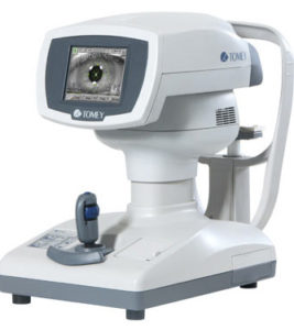
Tomey RT-7000
The Autorefractor Keratometer Topographer Tomey RT-7000 is a all-in-one unit. Patients are able to receive a comprehensive inspection without the hassle of moving from station to station. Refractometry, diameter measurement of cornea and pupil, keratometry, corneal topography, various color maps, CL fitting simulation.
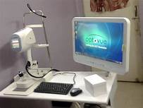
Optovue Ivue OCT
Optical coherence tomography (OCT) is a non-invasive imaging test. OCT uses light waves to take cross-section pictures of your retina. With an OCT we are able to see each of the retinas distinctive layers. We measure and map the thickness of each of your layers and that helps us with diagnosis. It also provides treatment guidance for glaucoma and diseases of the retina.
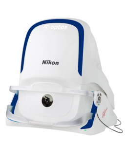
Optos California af

Getting an optomap image is fast, painless and comfortable. It is suitable for the whole entire family! To have the exam, you simply look into the device one eye at a time (like looking through a keyhole) and you will see a comfortable flash of light to let you know the image of your retina has been taken.
Under normal circumstances, dilation drops might not be necessary, but Dr. Kale will decide if your pupils need to be dilated depending on the health of your eyes. The image capture takes less than a half second and they are available immediately for you to see your own retina. You see exactly what your eye care practitioner sees – even in a 3D!
The benefits of having an optomap ultra-widefield retinal image taken are:
– optomap facilitates early protection from vision impairment or blindness.
– Early detection of life-threatening diseases like cancer, stroke, and cardiovascular disease.
The unique optomap ultra-widefield view helps your eye care practitioner detect early signs of retinal disease more effectively and efficiently than with regular eye exams.


